
lab manual for human anatomy and physiology
This foundational unit introduces the principles of human anatomy and physiology, exploring the structure and function of the body․ It includes key concepts, laboratory exercises, and virtual tools to enhance understanding of biological systems․
1․1 Key Concepts in Anatomy and Physiology
Key concepts in anatomy and physiology include understanding the levels of organization, from cells to systems, and the principles of homeostasis․ Anatomical terminology, such as directional terms and body planes, is essential for accurate communication․ The structure-function relationship highlights how body parts perform specific roles․ These foundational ideas are critical for navigating laboratory exercises and virtual dissections, providing a framework for exploring complex physiological processes in the human body․
1․2 Importance of Laboratory Work in Understanding Human Anatomy and Physiology
Laboratory work is crucial for applying theoretical knowledge in anatomy and physiology․ Hands-on exercises, such as dissections and virtual cadaver explorations, enhance spatial reasoning and practical skills․ These activities allow students to observe anatomical structures and physiological processes firsthand, fostering a deeper understanding․ Labs also encourage critical thinking and collaboration, preparing students for healthcare careers by bridging the gap between classroom learning and real-world applications․ This experiential approach ensures comprehensive learning and skill development․

Tissues
Tissues are groups of specialized cells that perform specific functions․ They form the building blocks of organs and systems, enabling the body to maintain homeostasis and function effectively․
2․1 Types of Tissues and Their Functions
Tissues are categorized into four primary types: epithelial, connective, muscle, and nervous․ Epithelial tissues form protective barriers and line body surfaces․ Connective tissues provide support and connect organs, while muscle tissues facilitate movement through contraction․ Nervous tissues transmit signals, enabling communication within the body․ Each tissue type plays a distinct role in maintaining overall physiological balance and organ function․
2․2 Epithelial, Connective, Muscle, and Nervous Tissues
Epithelial tissues form protective layers, such as skin and mucous membranes, and line internal organs․ Connective tissues provide structural support, connecting organs and forming bones, cartilage, and blood․ Muscle tissues enable movement through contraction, with types including skeletal, smooth, and cardiac muscles․ Nervous tissues specialize in conducting electrical impulses, comprising neurons and supportive cells like glial cells․ Together, these tissues form the building blocks of organs and systems, ensuring proper bodily functions․
Skeletal System
The skeletal system provides structural support, protects internal organs, and facilitates movement․ It comprises bones, joints, and ligaments, with functions including blood cell production and mineral storage․
3․1 Structure and Function of Bones
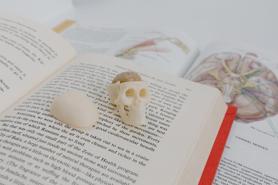
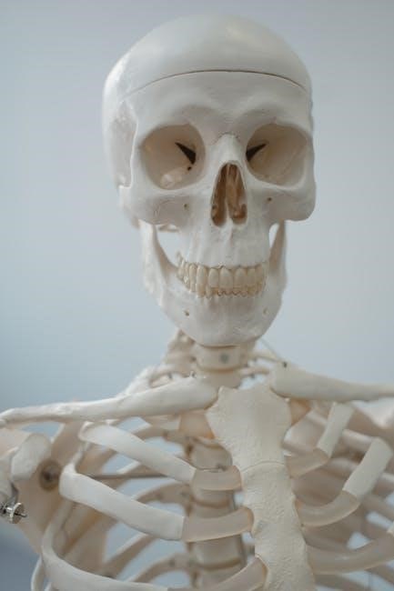
Bones are dynamic tissues composed of organic matrix (collagen) and inorganic minerals (calcium, phosphorus)․ The periosteum covers the outer surface, while the cortex provides strength and protection․ Spongy bone inside contains marrow, producing blood cells․ Bones support the body, protect organs, and facilitate movement․ Their structure includes compact and cancellous tissues, enabling functions like mineral storage and blood cell production․ Lab exercises explore bone anatomy using virtual cadaver tools and physical specimens․
3․2 Classification of Joints and Their Movements
Joints, or articulations, are classified based on their ability to move․ Synarthroses are immovable, like skull sutures․ Amphiarthroses allow limited movement, such as intervertebral discs․ Diarthroses are freely moving, enabling actions like flexion, extension, and rotation․ Examples include the shoulder (multidirectional) and knee (hinge)․ Virtual cadaver labs and physical models help students explore joint structures and their roles in movement, emphasizing the importance of joint classification in understanding human mobility and function․
3․3 Axial and Appendicular Skeleton
The axial skeleton includes the skull, vertebral column, ribs, and sternum, forming the body’s central framework․ The appendicular skeleton comprises the upper and lower limbs and their girdles, enabling movement․ Lab exercises, such as virtual cadaver dissections, allow students to explore these systems, understanding how bones articulate and function together․ These sessions emphasize the roles of axial bones in protection and appendicular bones in mobility, providing a comprehensive view of skeletal structure and function․
Muscular System
The muscular system comprises skeletal, smooth, and cardiac muscles, each with distinct functions․ Lab exercises focus on identifying major muscle groups and understanding their roles in contraction, movement, and maintaining body stability․
4․1 Types of Muscles and Their Functions
The muscular system includes three types of muscles: skeletal, smooth, and cardiac․ Skeletal muscles are voluntary, attached to bones, enabling movement and posture․ Smooth muscles are involuntary, found in internal organs, facilitating processes like digestion․ Cardiac muscle is specialized for the heart, ensuring rhythmic contractions․ Lab exercises involve identifying muscle types, understanding their roles, and exploring their physiological responses through dissections and virtual simulations․
4․2 Muscle Physiology and Movement
Muscle physiology involves the contraction and relaxation of muscle fibers, enabling movement and maintaining posture․ The sliding filament theory explains how actin and myosin interact to produce contractions․ ATP provides the energy for these processes․ Lab exercises include observing muscle contractions, analyzing EMG readings, and studying the role of the nervous system in controlling movement; Understanding muscle physiology is essential for grasping how the body executes voluntary and involuntary movements, from walking to heart contractions․
4․3 Major Muscle Groups and Their Actions
The human body contains over 600 muscles, grouped into major categories based on function and location․ The upper limb muscles, including the biceps and triceps, facilitate arm movements like flexion and extension․ Lower limb muscles, such as the quadriceps and hamstrings, enable walking and running․ The trunk muscles, including the abdominals and back muscles, support posture and spinal movements․ Facial muscles control expressions, while involuntary muscles manage functions like digestion; Lab exercises involve identifying and palpating these groups to understand their roles in movement and stability․
Nervous System
The nervous system is a complex network controlling communication, coordination, and bodily functions․ It consists of the central and peripheral nervous systems, enabling sensory input, processing, and motor responses․
5․1 Structure and Function of Neurons
Neurons, or nerve cells, are specialized cells designed for communication․ They consist of dendrites, a cell body, and an axon․ Dendrites receive signals, while the axon transmits them․ The cell body contains the nucleus and organelles essential for cell function․ Neurons communicate through electrical and chemical signals, enabling functions like movement, sensation, and thought․ Action potentials and synapses are key mechanisms in neuronal communication, allowing the nervous system to process and respond to information effectively․
5․2 Reflexes and Nerve Impulses
Reflexes are automatic responses to stimuli, involving a reflex arc․ Nerve impulses, or action potentials, are electrical and chemical signals that transmit information along neurons․ The process begins with stimulation of dendrites, leading to depolarization of the cell body․ If the threshold is reached, an action potential occurs, propagating along the axon․ Synapses transmit signals to other neurons or effector cells, enabling reflex actions and nervous system responses․ Lab exercises explore these mechanisms using simulations and physiological measurements․
5․3 Sensory and Motor Functions
Sensory functions involve the detection of stimuli, such as touch, pain, and temperature, through specialized receptors and neural pathways․ Motor functions enable voluntary and involuntary movements, controlled by the motor cortex, neurons, and muscle interactions․ Lab exercises, including nerve conduction studies and reflex testing, help students understand these processes․ Practical activities also explore how sensory input is processed and translated into motor responses, essential for coordinated body functions and maintaining homeostasis․
Circulatory and Respiratory Systems
The circulatory system transports oxygen and nutrients, while the respiratory system facilitates gas exchange․ Together, they maintain homeostasis, ensuring proper cellular function and overall health․
6․1 Blood Components and Their Roles
Blood is a vital fluid composed of plasma, red blood cells (RBCs), white blood cells (WBCs), and platelets․ Plasma, the liquid portion, transports nutrients and hormones․ RBCs, containing hemoglobin, carry oxygen throughout the body․ WBCs are essential for immune defense, fighting infections and diseases․ Platelets play a crucial role in blood clotting, preventing excessive bleeding․ Understanding these components and their functions is fundamental for studying circulatory health and disease diagnosis in laboratory settings․
6․2 Structure and Function of the Heart
The heart is a muscular organ with four chambers: the right and left atria, and the right and left ventricles․ Valves ensure blood flows in one direction․ The right side pumps blood to the lungs for oxygenation, while the left side distributes oxygenated blood to the body․ The heart’s electrical conduction system regulates contractions, maintaining a rhythmic heartbeat․ This structure enables the heart to efficiently circulate blood, supplying oxygen and nutrients to tissues and organs, essential for overall bodily function․
6․3 Mechanism of Breathing and Gas Exchange
Breathing involves the contraction and relaxation of the diaphragm and intercostal muscles, creating pressure changes that draw air into the lungs․ Gas exchange occurs in the alveoli, where oxygen diffuses into blood and carbon dioxide diffuses out․ This process is essential for delivering oxygen to tissues and removing carbon dioxide, maintaining proper cellular function and overall health․ Laboratory exercises often include simulations and measurements to visualize and understand these respiratory mechanisms effectively․
Digestive, Urinary, and Reproductive Systems
This section explores the digestive system’s role in nutrient absorption, the urinary system’s waste filtration process, and the reproductive system’s functions in gamete production and development․
7․1 Digestive System: Structure and Function
The digestive system processes food into nutrients for energy and growth․ It includes the mouth, esophagus, stomach, small intestine, and large intestine․ Mechanical digestion begins in the mouth, while the stomach uses gastric juices for chemical breakdown․ The small intestine absorbs nutrients into the bloodstream, and the large intestine manages water absorption and waste elimination․ Accessory organs like the liver, pancreas, and gallbladder support digestion by producing enzymes and bile, ensuring proper nutrient utilization․
7․2 Urinary System: Kidney Function and Urine Formation
The urinary system, comprising the kidneys, ureters, bladder, and urethra, filters blood to produce urine․ The kidneys, bean-shaped organs, contain nephrons that filter waste and excess substances․ Urine formation involves glomerular filtration, tubular reabsorption, and secretion․ The kidneys regulate water, salts, and pH, maintaining homeostasis․ Proper kidney function is essential for overall health, preventing toxin buildup and maintaining electrolyte balance effectively․
7․3 Reproductive System: Male and Female Anatomy
The reproductive system includes both male and female anatomical structures designed for gamete production and fertilization․ In males, the testes produce sperm, while the penis delivers semen․ In females, the ovaries release ova, and the uterus supports embryonic development․ Key components include the vas deferens, prostate, and seminal vesicles in males, and the fallopian tubes, cervix, and vagina in females․ These systems work together to enable reproduction and maintain species continuity through precise physiological processes․

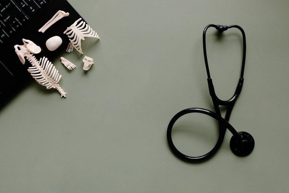
Laboratory Exercises
Laboratory exercises provide hands-on opportunities to explore human anatomy and physiology through dissections, observations, and interactive tools, enhancing understanding of complex biological systems and their functions․
8․1 Dissection and Virtual Cadaver Labs
Dissection and virtual cadaver labs provide hands-on experience for exploring human anatomy․ Physical dissections allow students to examine tissues and organs, while virtual cadaver tools like Anatomage offer 3D visualization․ These labs enable detailed study of internal structures, reinforcing anatomical knowledge․ Virtual platforms often include interactive features for labeling and layering tissues, enhancing learning․ Both methods ensure a safe and educational environment for students to practice and understand complex anatomical systems effectively․
8․2 Physiological Experiments and Measurements
Physiological experiments involve measuring bodily functions to understand system operations․ Students use tools like BIOPAC and BSL PRO to record data on heart rate, blood pressure, nerve impulses, and muscle contractions․ These exercises provide real-time insights into physiological processes, enabling students to correlate data with anatomical structures․ Hands-on activities, such as electrocardiograms and spirometry, enhance learning by demonstrating how body systems respond to stimuli and maintain homeostasis․ These experiments bridge theory and practice, fostering a deeper understanding of human physiology․
8;3 Microscopic Analysis of Tissues and Cells
Microscopic analysis enables students to study tissues and cells in detail, enhancing understanding of their structure and function․ Virtual cadaver dissection tools and histological slides are used to examine epithelial, connective, muscle, and nervous tissues․ Practical exercises include identifying cellular components and understanding tissue organization․ This hands-on approach helps students correlate microscopic observations with physiological processes, reinforcing concepts learned in anatomy and physiology labs․ These activities are essential for developing a strong foundation in histology and cellular biology․
Resources and References
Recommended lab manuals, textbooks, and online tools provide comprehensive guides for anatomy and physiology studies, offering interactive exercises, histological slides, and detailed illustrations for enhanced learning․
9․1 Recommended Lab Manuals and Textbooks
Elaine N․ Marieb’s Human Anatomy & Physiology Laboratory Manual is a top choice, featuring BIOPAC exercises and comprehensive guides for hands-on learning․ Terry R․ Martin’s Laboratory Manual for Human Anatomy & Physiology offers detailed dissection guides and interactive activities․ These resources, along with online tools like Anatomage virtual cadaver tables, provide students with immersive learning experiences․ They cater to healthcare-focused programs, emphasizing practical applications and visual aids for mastering complex anatomical structures․
9․2 Online Tools and Interactive Resources
Online tools like the Anatomage virtual cadaver table and BIOPAC exercises provide interactive learning experiences․ Digital platforms offer 3D models, simulations, and virtual dissections, enhancing understanding of anatomical structures․ These resources complement lab manuals, allowing students to explore complex physiological processes visually․ Interactive quizzes and multimedia content further engage learners, making anatomy and physiology more accessible and immersive for students in healthcare programs․